Home

Bien réplique district cornea slit lamp Aigle résultat Les notres

How to Use a Slit Lamp - American Academy of Ophthalmology

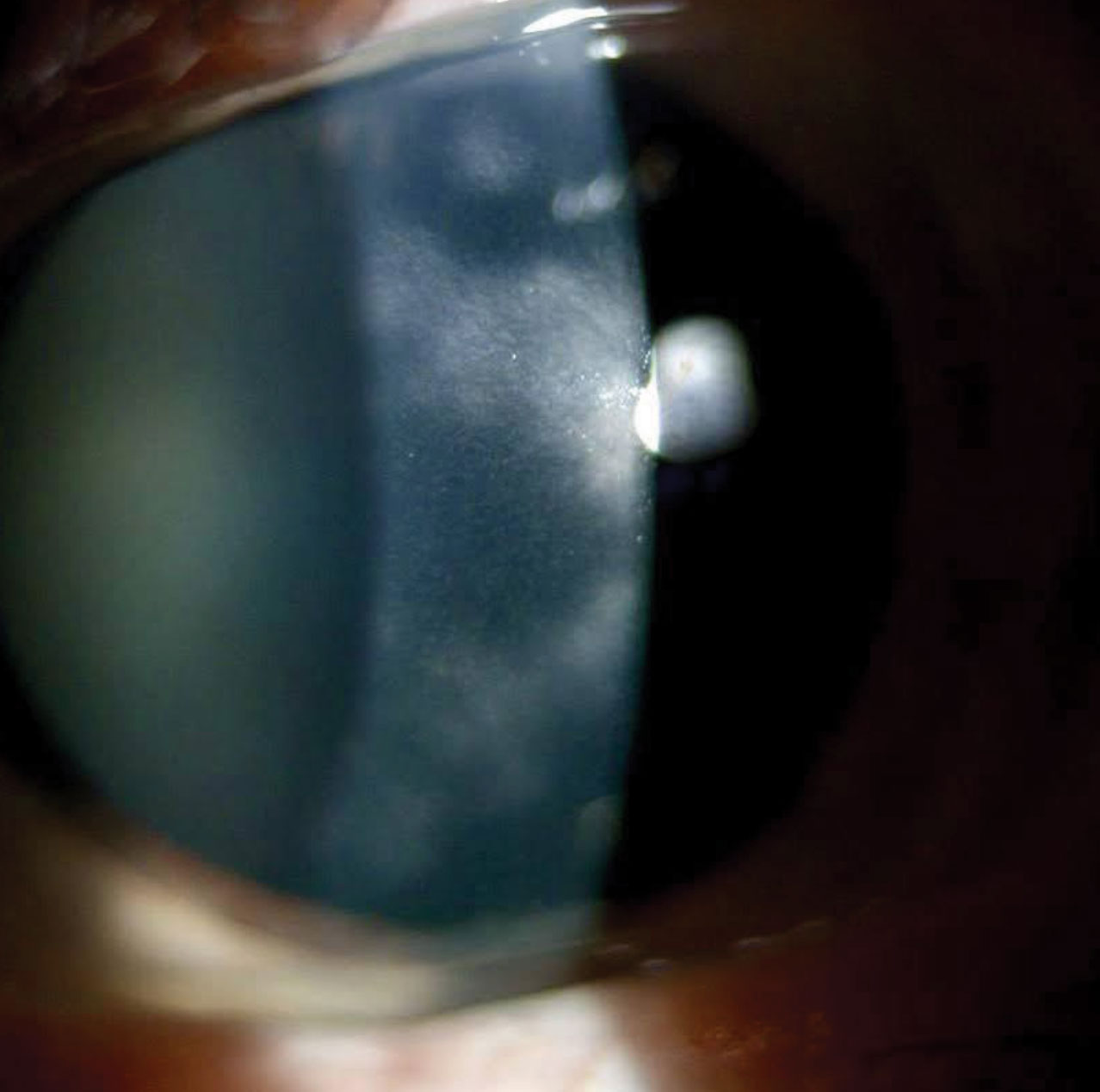
Post-LASIK Granular Corneal Dystrophy

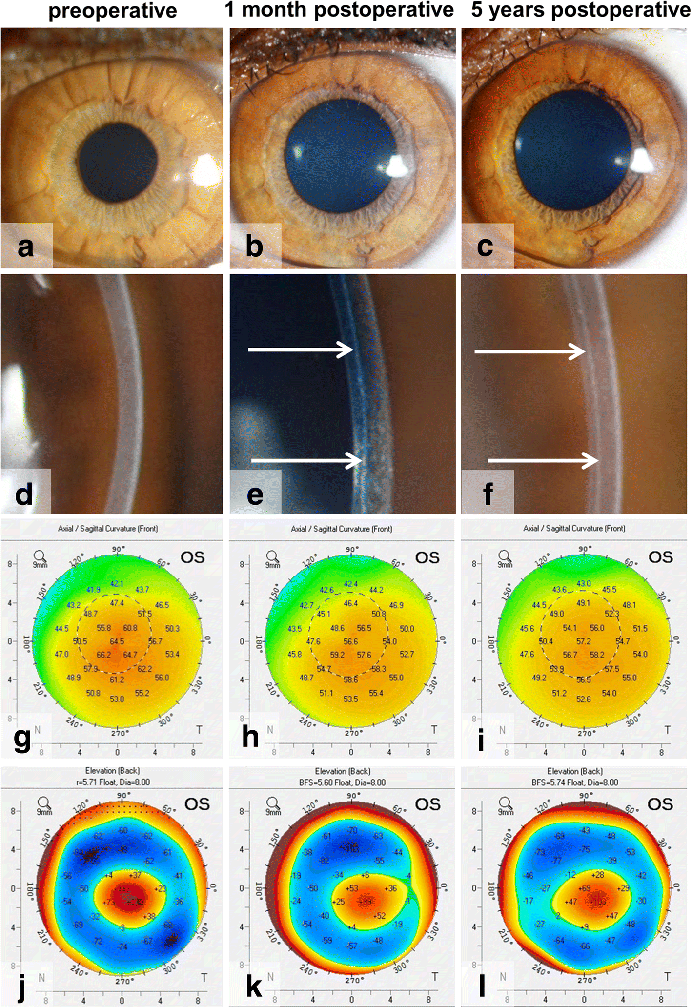
Bowman layer transplantation in the treatment of keratoconus | Eye and  Vision | Full Text
Bowman layer transplantation in the treatment of keratoconus | Eye and Vision | Full Text

Lecture: Using the Slit Lamp Microscope to Visualize the Ocular Structures  - YouTube
Lecture: Using the Slit Lamp Microscope to Visualize the Ocular Structures - YouTube

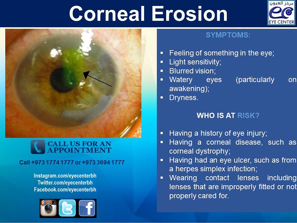
Eye Center by Dr. Ebtisam Al Alawi on X: "Corneal erosion affects the cornea,  the clear dome covering the front of the eye. If our #ophthalmologist  thinks you have corneal #erosion, we
Eye Center by Dr. Ebtisam Al Alawi on X: "Corneal erosion affects the cornea, the clear dome covering the front of the eye. If our #ophthalmologist thinks you have corneal #erosion, we
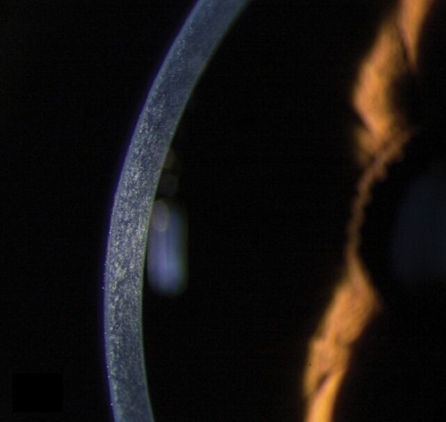
Cornea, Slit Lamp - Stock Image - C043/4137 - Science Photo Library

Cornea slit lamp biomicroscopy cross-section art digital art

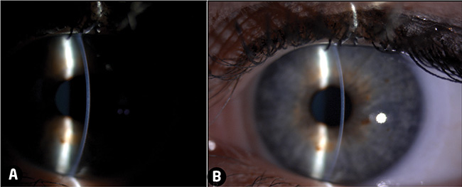
Slit-lamp Biomicroscopy

Fine-Tune Your Corneal Disease Diagnostic Skills

Slit lamp photography. In the arcuate beam from the slit lamp the... |  Download Scientific Diagram
Slit lamp photography. In the arcuate beam from the slit lamp the... | Download Scientific Diagram

Frontiers | Case report: A case of corneal deposits between binocular  descemet membrane and corneal endothelial layer after small-incision  lenticule extraction (SMILE) followed by HPV vaccine
Frontiers | Case report: A case of corneal deposits between binocular descemet membrane and corneal endothelial layer after small-incision lenticule extraction (SMILE) followed by HPV vaccine
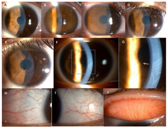
A, Slit-lamp biomicroscopy imaging of the cornea of this patient... |  Download Scientific Diagram
A, Slit-lamp biomicroscopy imaging of the cornea of this patient... | Download Scientific Diagram

JCM | Free Full-Text | Acute Foggy Corneal Epithelial Disease: Seeking  Clinical Features and Risk Factors
JCM | Free Full-Text | Acute Foggy Corneal Epithelial Disease: Seeking Clinical Features and Risk Factors
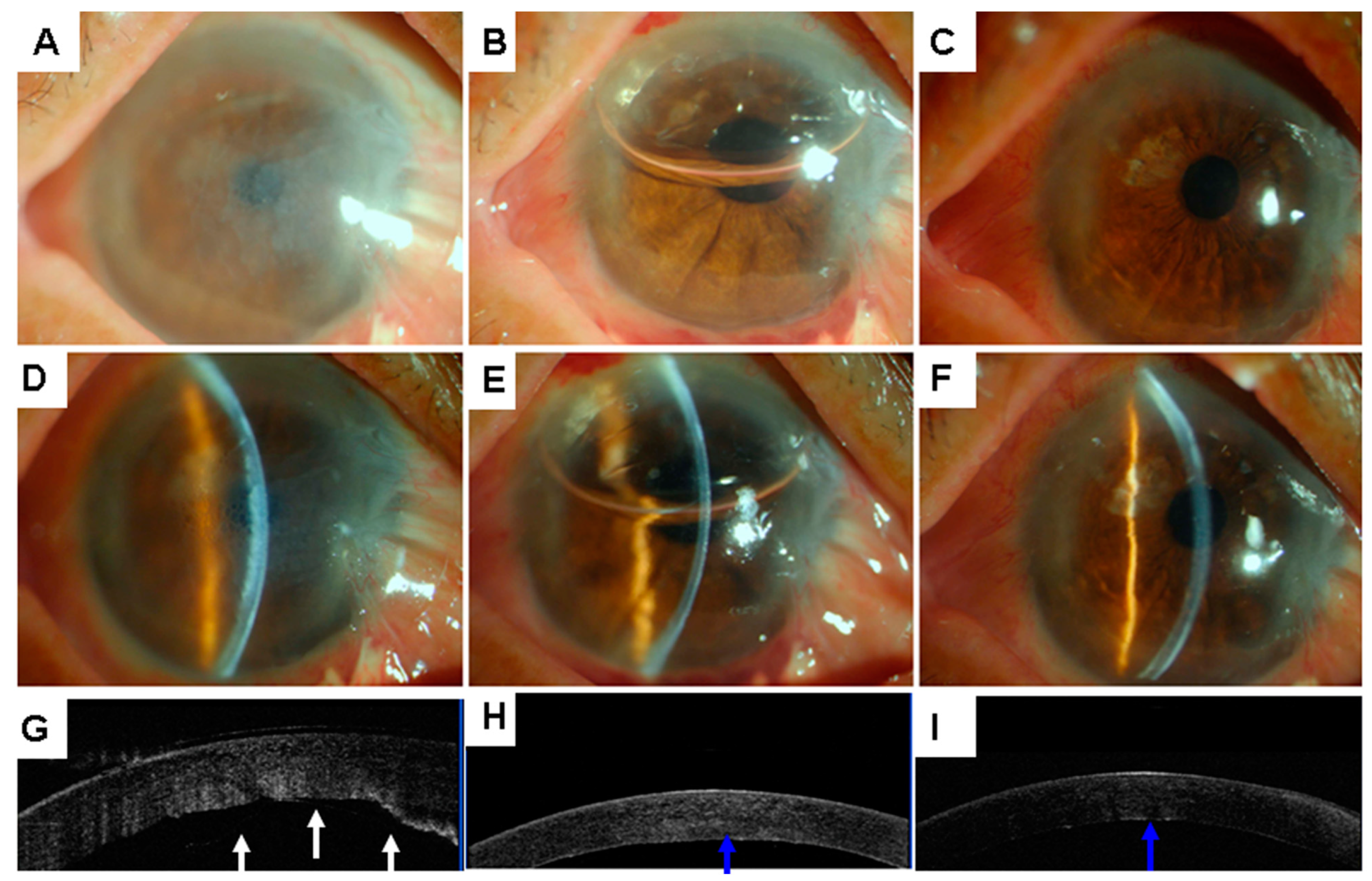
JCM | Free Full-Text | A Simple Repair Algorithm for Descemet’s  Membrane Detachment Performed at the Slit Lamp
JCM | Free Full-Text | A Simple Repair Algorithm for Descemet’s Membrane Detachment Performed at the Slit Lamp

Cornea and anterior eye assessment with slit lamp biomicroscopy, specular  microscopy, confocal microscopy, and ultrasound biomicroscopy | Semantic  Scholar
Cornea and anterior eye assessment with slit lamp biomicroscopy, specular microscopy, confocal microscopy, and ultrasound biomicroscopy | Semantic Scholar

Top tips for slit-lamp imaging - part 2

Ep 3: The Five (Possibly Six?) Layers of the Cornea — The Cornea Corner

Slit view on slit lamp examination showing a clear, flu | Open-i

Video slit lamp biomicroscope | Barnard Levit Optometrists

Corneal Pachymetry and Endothelial Microscopy by Slit-Lamp | IntechOpen

Slit Lamp Techniques: Optic Section and Parallepiped - YouTube

Fungal corneal ulcer (Video) - TimRoot.com

The following is a slit lamp photograph of a 60 year old woman with  complaints of chronic foreign body sensation. Which of the following is the  least likely cause of her condition?
The following is a slit lamp photograph of a 60 year old woman with complaints of chronic foreign body sensation. Which of the following is the least likely cause of her condition?
Corneal ulcer, slit-lamp examination - Stock Image - C034/8023 - Science  Photo Library
Corneal ulcer, slit-lamp examination - Stock Image - C034/8023 - Science Photo Library

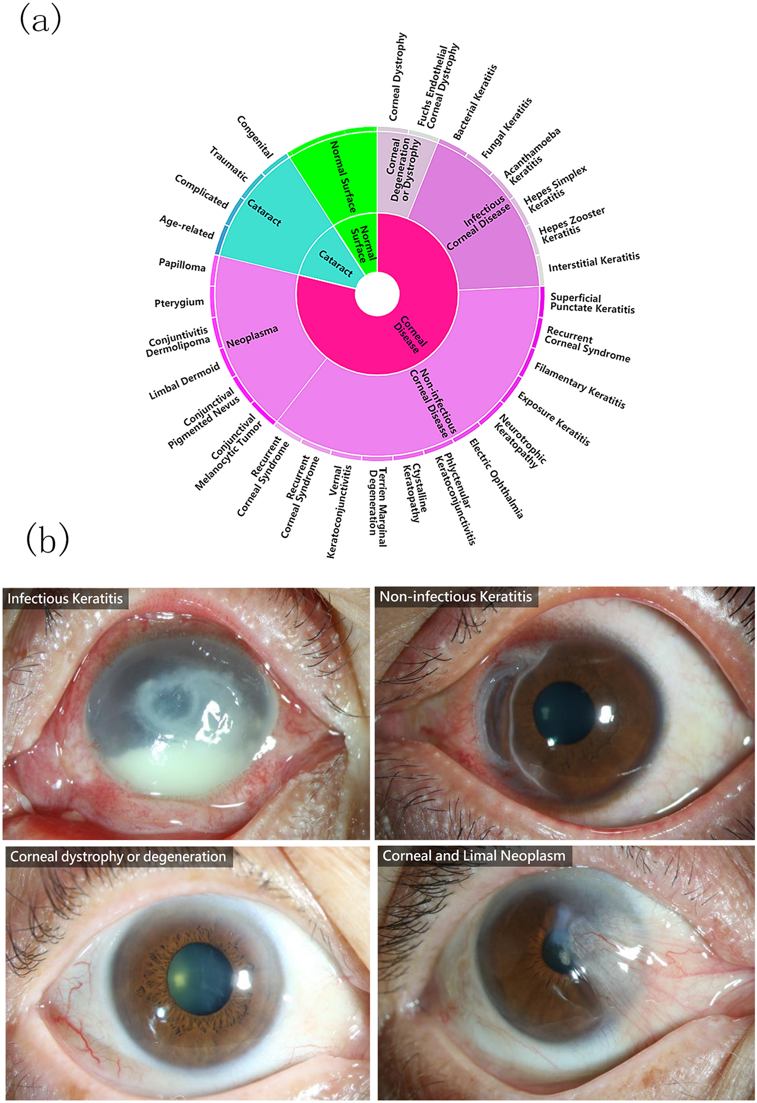
Deep learning for identifying corneal diseases from ocular surface slit-lamp  photographs | Scientific Reports
Deep learning for identifying corneal diseases from ocular surface slit-lamp photographs | Scientific Reports

Slit lamp technique #SPECULAR REFLECTION : Specular reflection is used to  analyze the corneal endothelial cells primarily to look for Fuch's  dystrophy,... | By Ophtalmology Lovers | Facebook
Slit lamp technique #SPECULAR REFLECTION : Specular reflection is used to analyze the corneal endothelial cells primarily to look for Fuch's dystrophy,... | By Ophtalmology Lovers | Facebook

Optician Online - CPD Archive